MRI in radiation therapy
The dose and beam can be accurately controlled with today’s modern LINACs to minimize patient risks.. The increasing precision of the linear accelerators requires a more precise irradiation planning. To determine the dose and the target position, the tumor and healthy tissue must be discriminated precisely from one another.
Magnetic Resonance Imaging (MRI) provides excellent soft tissue contrast for more detailed information to distinguish healthy tissue from gross tumor volume. In addition, physiological information such as diffusion and perfusion of the treatment region can be obtained to better define the target volume. In order to utilize the advantages of additional imaging by means of MRI in RT, a precise and reproducible patient position on both imaging modalities – CT and MRI – is essential for accurate image fusion later in the treatment chain.
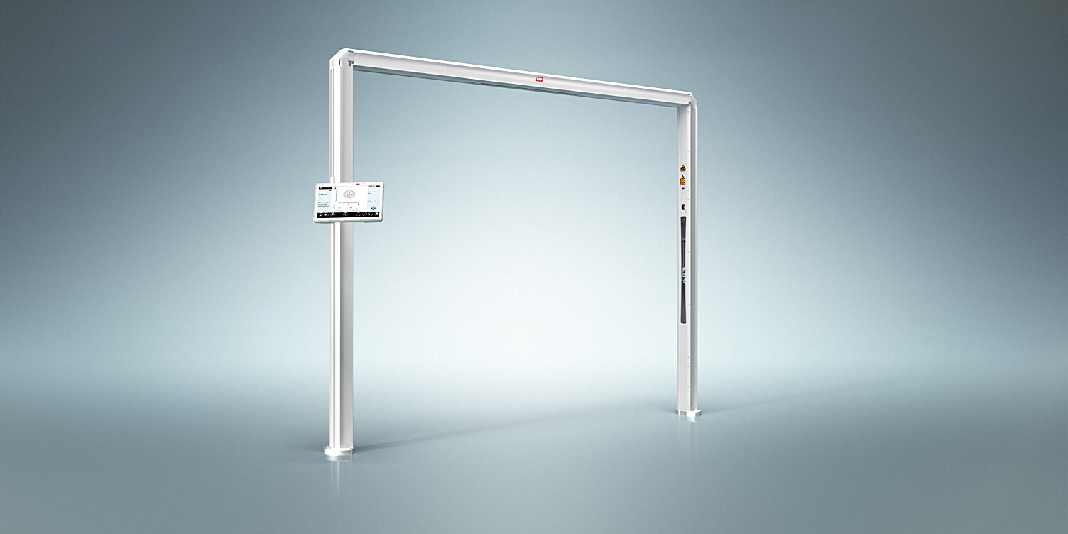
In an MR-only workflow, in the same way as for CT, an external laser system is required to project the patient-marking coordinates onto the patient’s skin.


